![]()
Part 1 Principles
1. Fluorescence microscope
2. Filterset
in FL-Mic
3. How concocal differs?
4.
What is confocal?
5.
Resolution in confocal
6. Optical
sectioning
7. Confocal image formation
and
time resolution
8. SNR in
confocal
9.
Variations of confocal
microscope
10. Special features from
Leica sp2 confocal
![]()
Part 2
Application
1. Introduction
2.
Tomographic view
(Microscopical CT)
3. Three-D reconstruction
4. Thick specimen
5. Physiological study
6.
Fluorescence detecting
General
consideration
Multi-channel detecting
Background correction
Cross-talk correction
Cross excitation
Cross emission
Unwanted FRET
![]()
Part
3 Operation and
Optimization
1.
Getting started
2. Settings in detail
Laser line
selection
Laser intensity and
AOTF control
Beam
splitter
PMT gain and offset
Scan
speed
Scan format, Zoom
and Resolution
Frame average, and
Frame accumulation
Pinhole and Z-resolution
Emission collecting rang
and Sequential scan
![]()
When Do
you need confocal?
FAQ
Are
you abusing
confocal?
Confocal Microscopy tutorial
Part 2 application of confocal microscopy
![]()
6. fluorescence detecting in confocal microscopy
Cross-talking 4: unwanted FRET
FRET (Fluorescence Resonance Energy Transfer) is a phenomena encountered when emission spectrum of one fluorophore falls into excitation spectrum of another fluorophore in the vicinity, when the two fluorophores are close enough, usually within 1-10 nm range, then the energy from emission of the first fluorophore will transfer to the second fluorophore and acts as excitation energy. The final effects will be a reduction of the first emission intensity and increase of the second emission intensity. There are many pairs of fluorophores with this property, naming a few: like FITC and TRITC, YFP and GFP, etc..
| The image here is taken from fluorescent beads which is
conjugated with both FITC and TRITC.
The unwanted FRET is hard to correct unless you reduce the intensity of first emission or choose other pair of fluorophore. As discussed above, unnecessarily mixing more fluorophores in the specimen can cause many adverse side-effects. |
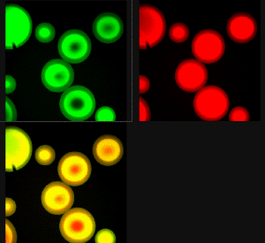 The
beads "should" show homogeneous green and red in the respective channel and
yellow in the overlay image because the two fluorophores are everywhere.
The
beads "should" show homogeneous green and red in the respective channel and
yellow in the overlay image because the two fluorophores are everywhere.
![]()
Statement about this web and
tutorial
For problems or questions regarding this web contact
e-mail:
This page was last updated 23.03.2004