![]()
Part 1 Principles
1. Fluorescence microscope
2. Filterset
in FL-Mic
3. How concocal differs?
4.
What is confocal?
5.
Resolution in confocal
6. Optical
sectioning
7. Confocal image formation
and
time resolution
8. SNR in
confocal
9.
Variations of confocal
microscope
10. Special features from
Leica sp2 confocal
Part 2
Application
1. Introduction
2.
Tomographic view
(Microscopical CT)
3. Three-D reconstruction
4. Thick specimen
5. Physiological study
6.
Fluorescence detecting
General
consideration
Multi-channel detecting
Background correction
Cross-talk correction
Cross excitation
Cross emission
Unwanted FRET
Part
3 Operation and
Optimization
1.
Getting started
2. Settings in detail
Laser line
selection
Laser intensity and
AOTF control
Beam
splitter
PMT gain and offset
Scan
speed
Scan format, Zoom
and Resolution
Frame average, and
Frame accumulation
Pinhole and Z-resolution
Emission collecting rang
and Sequential scan
When Do
you need confocal?
FAQ
Are
you abusing
confocal?
Confocal Microscopy tutorial
Part 1 Principles of Confocal microscopy
9. variations of confocal microscope
There are more than one way to achieve confocal effect. It doesn't matter if a system use laser, pinhole or not, if the multiplication of two PFS can be achieved, so does the confocal effect. There are three major types of confocal system available on the market.
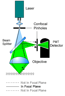
Laser scanning confocal Microscope (LSCM)
 The archetype of confocal microscope in the confocal
family. It utilizes laser and illumination pinhole to get point-like light
source illumination on the specimen. In recent model by most manufacturer, the
lasers are usually controlled by AOTF (acousto-optical tunable filter) for fast,
real time change of laser line and intensity. Detecting pinhole is used to
get rid of out-of-focus signal. PMTs are used as detecting
device.
The main advantage of this type is its combination of good image quality, versatile functionality and reliability and easy to maintain. 6 visible laser channel, 1 UV laser channel, 2 infra red laser channel can be fitted into the system and enable user to detecting up to eight channels simultaneously.
The disadvantage of this type is its slow frame rate, its photo bleaching and photo-cytotoxicity due to the indiscriminately illumination and excitation, exciting on the full length of the specimen (although only focal-plane signal is collected by PMT).
multiphoton laser confocal microscope (MP)
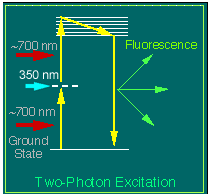
  This type of confocal uses two lasers for a single
excitation. It is based on the phenomena that two high-energy lasers of
longer wavelength focused on the single point will produce a shorter excitation
at the half of their original wavelength. For example, two 700 nm infra-red laser will
produces about 350 nm UV excitation. Since its high energy demanding, only pulsed
laser can meet its requirement. Two point illumination itself provide two PFS, detecting pinhole is not need in this configuration.
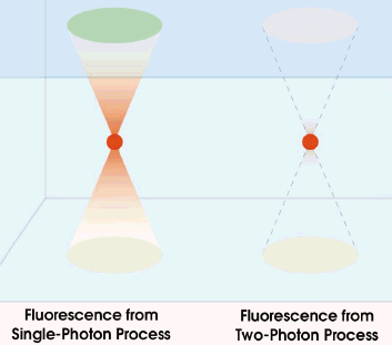
As shown in figure on the right side, the excitation of fluorophore occurs only at the
focal point where two laser meet, (while in single photon system, the excitation
is on the whole illuminated length, although only emission from focal point is
detected). This type of confocal uses two lasers for a single
excitation. It is based on the phenomena that two high-energy lasers of
longer wavelength focused on the single point will produce a shorter excitation
at the half of their original wavelength. For example, two 700 nm infra-red laser will
produces about 350 nm UV excitation. Since its high energy demanding, only pulsed
laser can meet its requirement. Two point illumination itself provide two PFS, detecting pinhole is not need in this configuration.
As shown in figure on the right side, the excitation of fluorophore occurs only at the
focal point where two laser meet, (while in single photon system, the excitation
is on the whole illuminated length, although only emission from focal point is
detected). |
The main advantage of MP is its low photo-cytotoxicity, since the long wavelength infra red laser has less toxicity than short wavelength. It also bleaches less than LSCM because only the focal point is affected. For this reason, MP suits living cell imaging, point bleach experiment, and other physiologic studies well. Since long wave length pulse laser has less energy loss when penetrating specimen, MP can work with even thicker specimen than single photon confocal.
The disadvantage of MP is the high price for building-up and maintaining the system. The image quality is a little bit deteriorated since there is no pinhole.
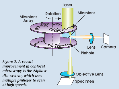
Disk scanning confocal microscope (nipkow disk system)
 In this system,
a spinning disk with multiple small hole is installed between the light source and specimen to shed point-like source on
specimen. The same hole serves as detecting pinhole to remove out-of-focus
light. Instead of PMT, Nipkow system use CCD camera as detector. Since the point
light source is provided by disk, laser is not necessary and visible light can
be used which reduces the photo-toxicity. But in practice, laser is still
commonly used in Nipkow system to get enough intensity.
The main advantage of Nipkow system is its high scan speed. The scanning speed of the disk can reach 300 frame per second, but the high scanning speed of disk is compromised by low readout speed of CCD camera as discussed in section 7. Ironically, the high "readout" speed of PMT in LSCM system is compromised by the slow scanner speed.
The disadvantage of Nipkow system is its relative poor image quality, limitation on specimen thickness, and only one detecting channel can be used at a time. Sequential detecting has to be used for multi-channel then the high speed advantage of the system lost.
Statement about this web and
tutorial
For problems or questions regarding this web contact
e-mail:
This page was last updated 23.03.2004