![]()
Part 1 Principles
1. Fluorescence microscope
2. Filterset
in FL-Mic
3. How concocal differs?
4.
What is confocal?
5.
Resolution in confocal
6. Optical
sectioning
7. Confocal image formation
and
time resolution
8. SNR in
confocal
9.
Variations of confocal
microscope
10. Special features from
Leica sp2 confocal
![]()
Part 2
Application
1. Introduction
2.
Tomographic view
(Microscopical CT)
3. Three-D reconstruction
4. Thick specimen
5. Physiological study
6.
Fluorescence detecting
General
consideration
Multi-channel detecting
Background correction
Cross-talk correction
Cross excitation
Cross emission
Unwanted FRET
![]()
Part
3 Operation and
Optimization
1.
Getting started
2. Settings in detail
Laser line
selection
Laser intensity and
AOTF control
Beam
splitter
PMT gain and offset
Scan
speed
Scan format, Zoom
and Resolution
Frame average, and
Frame accumulation
Pinhole and Z-resolution
Emission collecting rang
and Sequential scan
![]()
When Do
you need confocal?
FAQ
Are
you abusing
confocal?
Confocal Microscopy tutorial
Part 1 Principles of Confocal microscopy
![]()
5. Lateral and Axial Resolution in confocal system
Point Spread Function (PSF) and resolution
 In optical lens system,
lens can magnify an object because of its geometric optical property. But because of the wave property of light, diffraction
occurs when light pass it like the depicted in the figure. The image formed by
a point light at
the focal plane is not a single point, instead, the point distributed in a
possible area on the image plane. This distribution has higher probability
(higher intensity) at center and lower probability (lower intensity) at
peripheral, i.e., the point is spread after lens. This is Point Spread Function
(PSF). PSF describes the probability for an area where a given point will appear. In optical lens system,
lens can magnify an object because of its geometric optical property. But because of the wave property of light, diffraction
occurs when light pass it like the depicted in the figure. The image formed by
a point light at
the focal plane is not a single point, instead, the point distributed in a
possible area on the image plane. This distribution has higher probability
(higher intensity) at center and lower probability (lower intensity) at
peripheral, i.e., the point is spread after lens. This is Point Spread Function
(PSF). PSF describes the probability for an area where a given point will appear.The distribution pattern looks like a disc with a central high intensity spot and many concentric rings, a pattern called Airy disc. Airy pattern describes the light intensity distribution as a function of distance from the optical axis.
|
 The intensity distribution takes
about 84%, 91%, 94% total photons within the inner, the 2nd and the third ring,
and weaker further along distance. The intensity distribution takes
about 84%, 91%, 94% total photons within the inner, the 2nd and the third ring,
and weaker further along distance.The optical resolution of a lens is determined by the size of the inner dark ring of its Airy disk. The radius of the inner ring is related to the aperture of the lens. The smaller the aperture, the larger the PSF and the bigger the inner ring, thus the lower its optical resolution. When single point resolution is concerned, the radius of the inner ring, as shown in the figure, is used to define the resolution, thus called resolution element (resel). When point-to-point resolution is concerned, the resolution is defined as the distance of the two points where the maximum of one point is just above the first minimum of the second point (Rayleigh criterion). |
Another frequently used definition for resolution of a lens is the Full Width of Half Maximum (FWHM) of the airy disk which is diameter of the central spot where the intensity drop to its half maximum. FWHM is widely used because it is easy to measure.
| The diffraction
depends on wavelength of the light in use, the numerical aperture (NA) of
the lens. Formula 1 is used for the lateral resolution: |
|
| In confocal microscope, due to its improved contrast, there
is a slight gain of resolution but it is still diffraction limited. When pinhole is infinite close to zero, (in
practical, < 0.25 AU), lateral Res. is calculated by Formula 2: Compare these two formula, you can see that, in theory, confocal system provides higher resolution than conventional. It comes from the ratio of (0.51/0.37) and λem/λex, roughly, about 1.4 times of conventional. |
But the pinhole size can not be infinite to zero, when pinhole is between 0.25-1 AU, a value between 0.37-0.51 is used to replace 0.37 in the formula. When pinhole is over 1 AU, 0.51 has to be used and the resolution advantage over conventional fluorescence microscope is minimized to the ratio of Em/Ex: in case of FITC, it is 510/488 = 1.05 times.
For pinhole < 0,25 AU, the resolution advantage is often compromised by the vulnerability to noise due to its reduced detecting volume and general weaker signal and deteriorated SNR. For detailed discussion, see section 9, SNR in confocal.
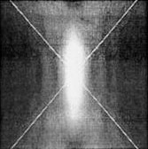 Point spread
function applies not only to the focal plane horizontally, but also to the
plane vertically. A perpendicular cut of the Airy disc shows a even
wide spread range. That is why the Z-resolution of a lens is
worse than its lateral resolution. Point spread
function applies not only to the focal plane horizontally, but also to the
plane vertically. A perpendicular cut of the Airy disc shows a even
wide spread range. That is why the Z-resolution of a lens is
worse than its lateral resolution.When pinhole is close to zero (or < 0,25 AU in practice): Formula 3 :  is used for
calculation. is used for
calculation.In general, the Z-Resolution is twice of the x-y-Resolution. 0.64-0.88 is used to replace 0.64 when pinhole is at 0.25-1.0 AU. |
The effect of PSF makes image from any diffraction-limited system blurred and distorted, or convoluted (twisted). Deconvolution algorithm has to be used to correct this distortion after data collection if high demanding of a true representative of original is required.
![]()
Statement about this web and
tutorial
For problems or questions regarding this web contact
e-mail:
This page was last updated
23.03.2004