![]()
Part 1 Principles
1. Fluorescence microscope
2. Filterset
in FL-Mic
3. How concocal differs?
4.
What is confocal?
5.
Resolution in confocal
6. Optical
sectioning
7. Confocal image formation
and
time resolution
8. SNR in
confocal
9.
Variations of confocal
microscope
10. Special features from
Leica sp2 confocal
![]()
Part 2
Application
1. Introduction
2.
Tomographic view
(Microscopical CT)
3. Three-D reconstruction
4. Thick specimen
5. Physiological study
6.
Fluorescence detecting
General
consideration
Multi-channel detecting
Background correction
Cross-talk correction
Cross excitation
Cross emission
Unwanted FRET
![]()
Part
3 Operation and
Optimization
1.
Getting started
2. Settings in detail
Laser line
selection
Laser intensity and
AOTF control
Beam
splitter
PMT gain and offset
Scan
speed
Scan format, Zoom
and Resolution
Frame average, and
Frame accumulation
Pinhole and Z-resolution
Emission collecting rang
and Sequential scan
![]()
When Do
you need confocal?
FAQ
Are
you abusing
confocal?
Confocal Microscopy tutorial
Part 2 application of confocal microscopy
![]()
2. Tomographic view of specimen
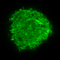
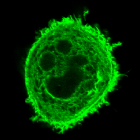
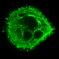
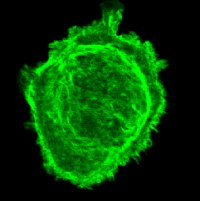
The depth discrimination feature of confocal microscopy enables user to look specimen along certain axis, usually Z-axis, slice by slice at a chosen distance interval to reveal the structural differences or fluorescence distribution differences inside the specimen. Somehow like a Röntgenist examines patients with CT: "computerized X-ray tomography", the tomography of a cell or thick tissue can be obtained in the similar way. That is why people sometimes refer this as "Microscopic CT", although not accurate as a full description for all the function of confocal microscopy, it is very suitable for this application. This function is very useful especially when you think the spatial distribution of the structures make sense in addition to its positive staining, even if you are not interested to get a Z-series for 3D reconstruction at all.
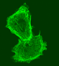
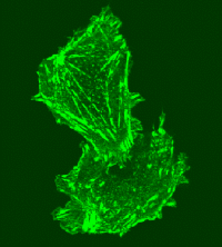
The four pictures below are taken from the same cell at different Z-position from top to bottom untill the cell attachment plane. It reveals differences in both internal structures or cell surface features among optical sections which can not be seen in normal wide field microscopy.
 |
 |
 |
 |
Not just the structure of a single cell, the relationship of adjacent cells at different plane can also be viewed clearly like below:
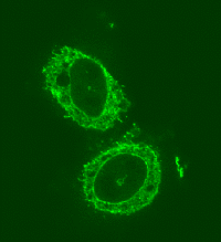 |
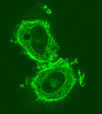 |
 |
 |
This technique can also be used to differentiate
whether a diffused staining is membrane staining and pan-cytoplasm staining.
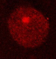
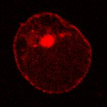
The left picture shows a staining pattern which is difficult to tell its nature.
Cutting it deeper along Z-axis reveals a
circular profile which enables you say with sure this is a membrane staining.
![]()
Statement about this web and tutorial
For problems or questions regarding this web contact
e-mail:
This page was last updated
23.03.2004