Advanced
Microscopy unit
Department
of Pathology
Haartman
Institute, University of Helsinki
![]()
Facilities in AMU
Electron Microscopy:
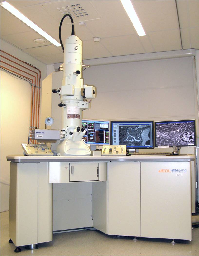
JEOL 1400 Transmission Electron Microscope
Location: Room DK 139, Haartman Institute
|
|
Accelerate Voltage Resolution Magnification Digital Camera Film Camera Stage Tilting range |
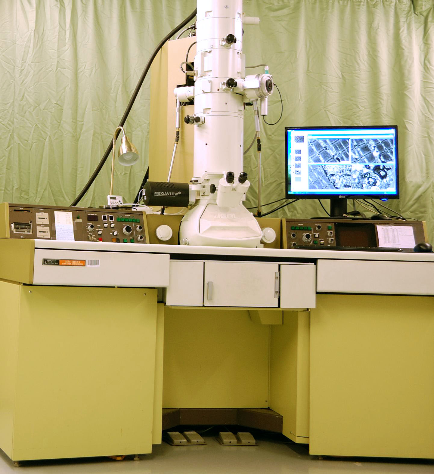
JEOL 1200 EX Transmission electron microscope
Location: Room EK 247, Haartman Institute
|
|
Acceleration Voltage Resolution Magnification Digital Camera Film Camera Stage: manual |
|
|
|
|
|
Gun
and filament: Accelerate voltage 0.2-30 KV Resolution Magnification Detectors Stage: motorized Tilting range: -15-75 |
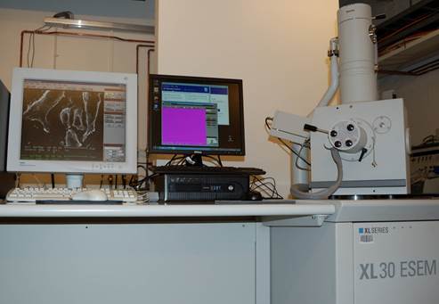
FEI XL-30 ESEM (Environmental Scanning Electron Microscope)
Location: Room EK 247, Haartman Institute
Auxiliary facilities for electron microcopy
Location: Room EK 247, Haartman Institute
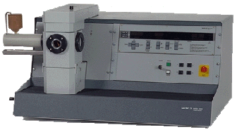
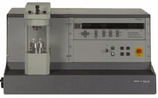
Balzer MED 020 multiple function,
modular coating device
For high resolution coating of EM specimen and grids supporting
film.
When different modules (chambers and top flanges) are used like showing below,
it can be used as different function.
|
|
Carbon Coating: thread
and E-gun rod coating. |
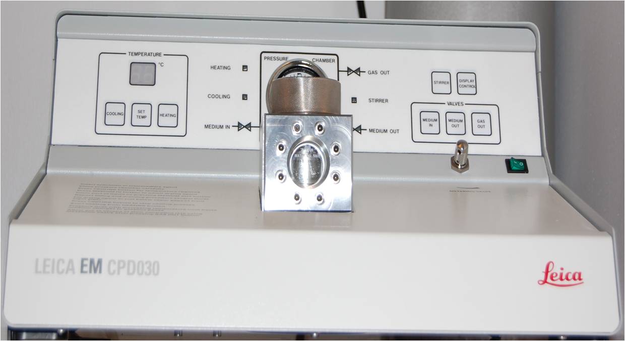
Leica EM CDP 030 critical point drier for specimen preparation of both
TEM and SEM.
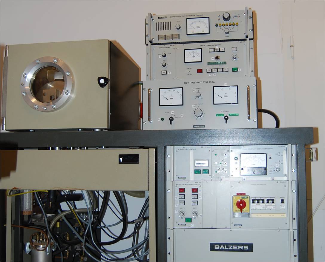
Freeze Fracture device Balzers BAF 400 D for making freeze fracture
and simultaneously surface coating with metal, carbon or both.
specimen preparation Lab
Location: Room DK 138, Haartman Institute
For tissue embedding, ultra thin
sectioning, staining, etc., a specimen preparation lab is available. Equipped
with following devices:
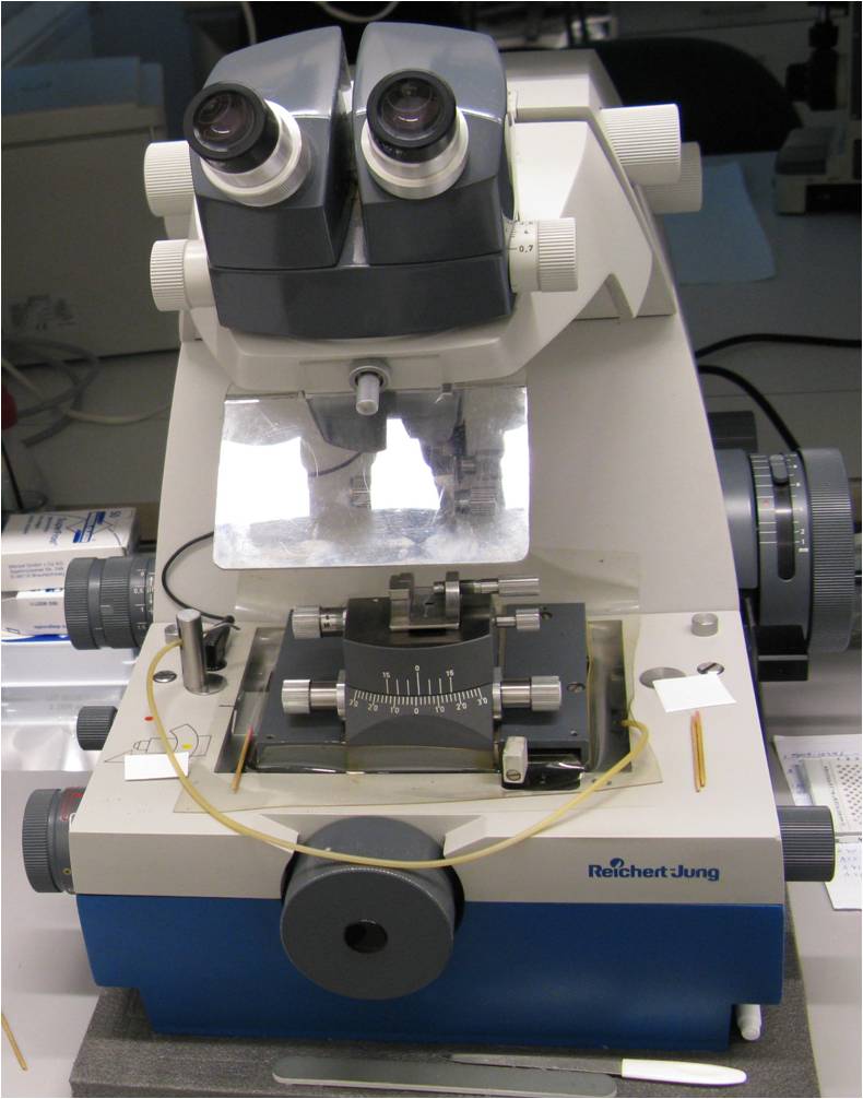
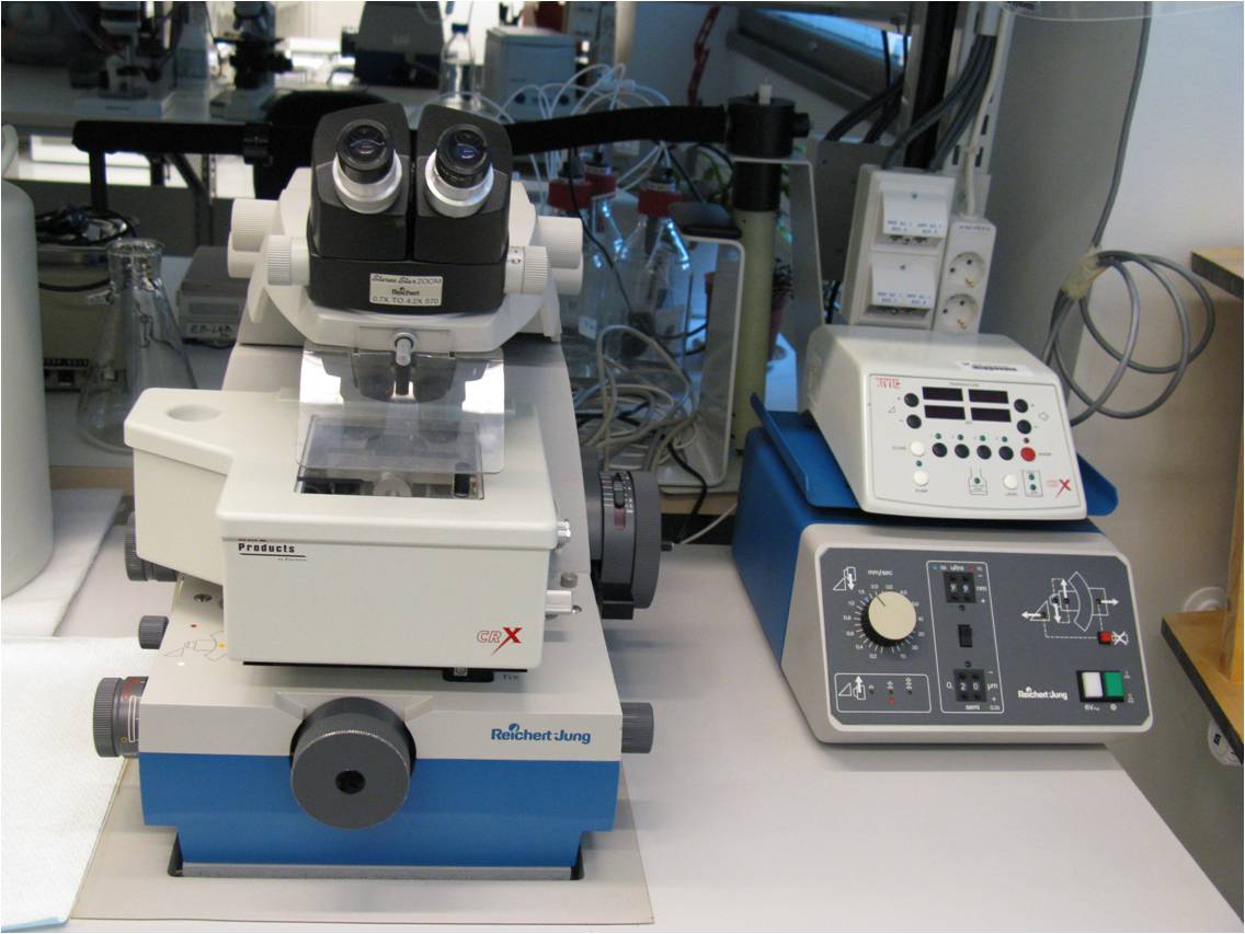
Ultra-microtome
|
Reichert-Jung ultra microtome for
routing ultra thin section |
Reichert-Jung ultra microtome with
cryo-box for frozen untra-thin sectioning |
|
|
|
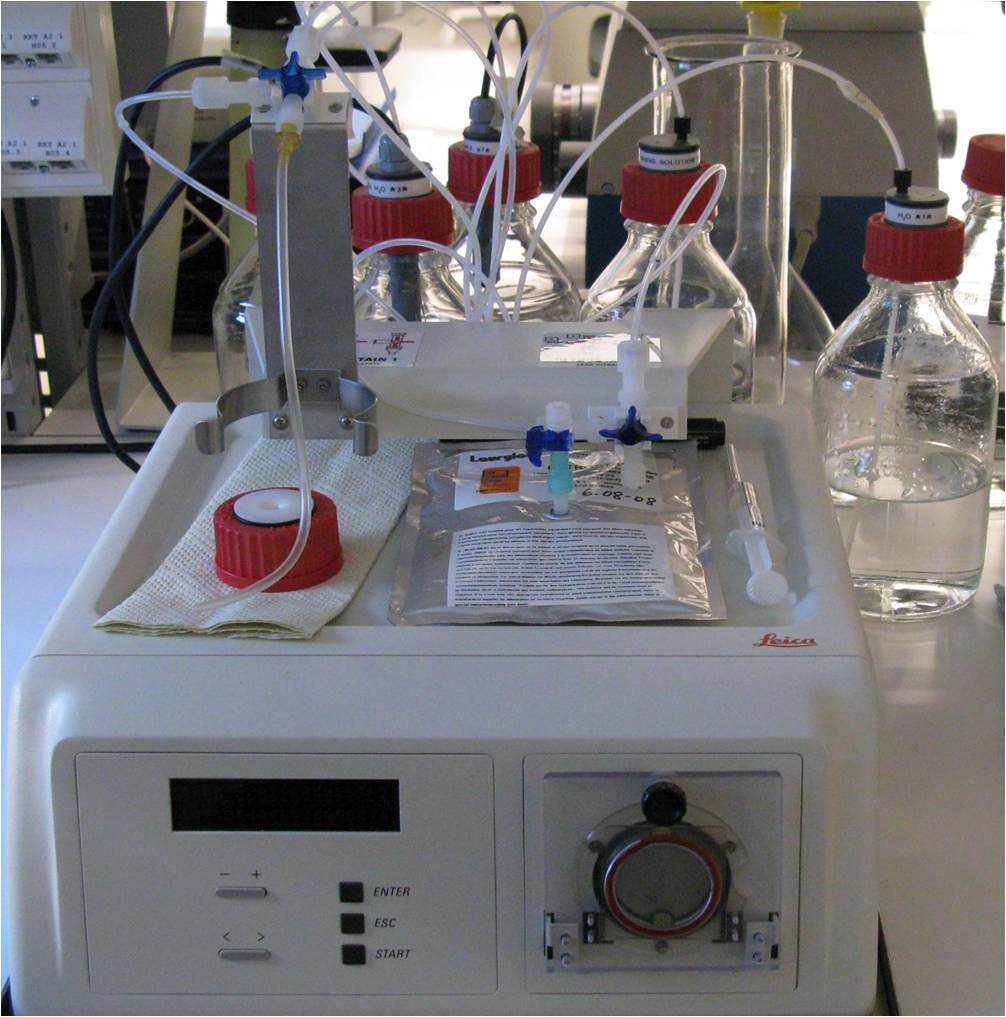
Leica EMStain for automatic thin section
staining
Next ![]()
![]()
Statement about this web and tutorial
For problems or questions regarding this web contact webmaster
This page was last updated
08.12.2009