![]()
Part 1 Principles
1. Fluorescence microscope
2. Filterset
in FL-Mic
3. How concocal differs?
4.
What is confocal?
5.
Resolution in confocal
6. Optical
sectioning
7. Confocal image formation
and
time resolution
8. SNR in
confocal
9.
Variations of confocal
microscope
10. Special features from
Leica sp2 confocal
Part 2
Application
1. Introduction
2.
Tomographic view
(Microscopical CT)
3. Three-D reconstruction
4. Thick specimen
5. Physiological study
6.
Fluorescence detecting
General
consideration
Multi-channel detecting
Background correction
Cross-talk correction
Cross excitation
Cross emission
Unwanted FRET
Part
3 Operation and
Optimization
1.
Getting started
2. Settings in detail
Laser line
selection
Laser intensity and
AOTF control
Beam
splitter
PMT gain and offset
Scan
speed
Scan format, Zoom
and Resolution
Frame average, and
Frame accumulation
Pinhole and Z-resolution
Emission collecting rang
and Sequential scan
When Do
you need confocal?
FAQ
Are
you abusing
confocal?
Confocal Microscopy tutorial
Part 2 application of confocal microscopy
4. Thick specimen
As emphasized in previous section, one forte of confocal microscopy is its power to resolve thick specimen thanks to its depth discrimination property. It can be used for tissue block, small organ, embryo, etc.
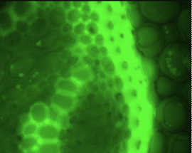
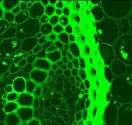
The following two images are taken from the same field of the plant of 60 Ám thick by different means. The first one is taken by CCD camera in the wild field microscope configuration. the second one is taken by confocal microscopy with pinhole size at one Airy unit. The difference on the results is striking.
 |
 |
It is worth to mention that the difference is not just the sharpness or crisp of the images, but also the ability of resolving the internal structures. Many fine structures clearly visible in confocal image are obscured or totally invisible in the image taken under wild field microscope.
For even thicker specimen, no satisfied image can be obtained at all.
Statement about this web and
tutorial
For problems or questions regarding this web contact
e-mail:
This page was last updated
23.03.2004